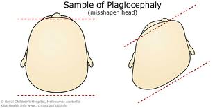
Positional plagiocephaly (play-gee-oh-seff-a-lee) is the term used to describe a flattened or misshapen head that may result from crowding within the womb or from an infant being placed in the same position (such as on the back) for long periods of time.
Health care providers also use the term brachycephaly (bray-kee-seff-a-lee) to describe the flattening of the back of the skull. Positional plagiocephaly and brachycephaly often occur together.
The general term plagiocephaly refers to a head that is abnormally shaped from a variety of causes. Positional plagiocephaly is only one type of plagiocephaly. Other types can result from: skull sutures closing too soon (called craniosynostosis); twisted neck present at birth (called congenital muscular torticolis); and fluid around the brain (called hydrocephaly). Plagiocephaly can sometimes be severe and may require surgical treatments.
What causes positional plagiocephaly?
Positional plagiocephaly usually results from an infant being placed in the same position, such as on the back, for long periods of time.
Babies' skulls are soft and are made up of several skull plates. These movable plates have space between them, called sutures, that allow the head to be flexible so that the brain can grow. If the head is left in the same position for long periods of time, the plates move in a way that leaves a flat spot.
The most common causes of positional plagiocephaly include:
- Too little time spent upright—sometimes called "cuddle" time
- Too little "Tummy Time" when the baby is awake and supervised
- Too much time in car seats, carriers, and bouncy seats
How can I help prevent positional plagiocephaly?
Most cases of positional plagiocephaly can be prevented (and sometimes corrected) by repositioning, which relieves pressure from the back of an infant’s head. Techniques for repositioning include:
- Providing "Tummy Time" when your baby is awake and someone is watching. Tummy Time not only helps prevent flat spots, but it also helps the baby's head, neck, and shoulder muscles get stronger as part of normal development. Parents and caregivers can try Tummy Time 2 or 3 times a day, for short periods of time, until the baby gets used to being on the tummy. Once the baby begins to enjoy the position, parents can try longer periods of time or increase frequency of Tummy Time. [link to http://www.nichd.nih.gov/health/topics/Tummy_Time.cfm]
- Changing the direction that your baby lies in the crib from one week to the next. For example, have the baby's feet point toward one end of the crib for a few days, and then change the position so his or her feet point toward the other end of the crib. This change will encourage the baby to turn his or her head in different directions to avoid resting in the same position all the time.
- Avoiding too much time in car seats, carriers, and bouncers while the infant is awake.
- Getting "cuddle time" with the baby by holding him or her upright over one shoulder often during the day.
- Changing the location of the baby's crib in the room so that he or she has to look in different directions to see the door or the window.
What are the treatments for positional plagiocephaly?
In most cases, flat spots on the head go away on their own once an infant starts sitting up and crawling. Repositioning (described above) can also successfully correct positional plagiocephaly.
If the problem is detected early, more severe positional plagiocephaly can usually be treated successfully under a health care provider's direction. Some options for this treatment include:
If the abnormal shape is the result of some other disorder, and is not related to position, a health care provider may also recommend helmets and bands. Or, if the head is severely misshapen or the result of craniosynostosis, surgery may be required to correct the problem.
Talk to your health care provider about your baby’s head shape at each well-baby checkup.
Source: Eunice Kennedy Shriver | Nat'l Institute of Child Health and Human Development
If the problem is detected early, more severe positional plagiocephaly can usually be treated successfully under a health care provider's direction. Some options for this treatment include:
- A custom helmet that prevents the baby's head from always falling to one side when lying down. Most importantly, it allows the baby’s skull to expand into a more rounded shape as the brain and skull grow.
- Custom bands can also be used to help reshape a baby's head. Bands place a small amount of pressure on the baby's skull in the areas that need reshaping. A health care provider adjusts the band regularly as the baby’s head grows.
If the abnormal shape is the result of some other disorder, and is not related to position, a health care provider may also recommend helmets and bands. Or, if the head is severely misshapen or the result of craniosynostosis, surgery may be required to correct the problem.
Talk to your health care provider about your baby’s head shape at each well-baby checkup.
Source: Eunice Kennedy Shriver | Nat'l Institute of Child Health and Human Development
If you would like more information on Plagiocephaly, Torticollis or Cranial Remolding Helmets, please feel free to conact us here.


Gary Tate Jr.
President and Founder
Alexandra's P.H.A.T.E.
Pittsburgh, PA
412-860-4557
http://draft.blogger.com/gary@alexandrasphate.org
President and Founder
Alexandra's P.H.A.T.E.
Pittsburgh, PA
412-860-4557
http://draft.blogger.com/gary@alexandrasphate.org